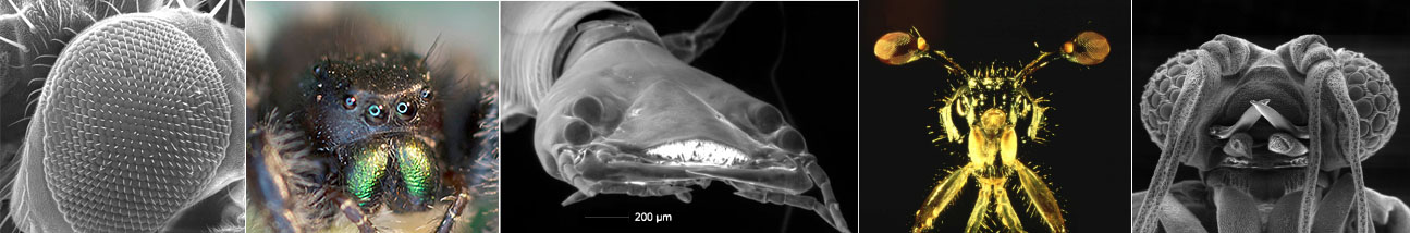
Invertebrate eyes are frequently highly specialized and optimized to perform a particular visual task. When we establish exactly what they are particularly good at, how they are adapted to this task, and what they are specialized for, we can often uncover functional designs that nobody has thought of thus far. For example, some of our work on a little parasite, Xenos peckii (of the order Strepsiptera) has inspired engineers to design highly successful micro-cameras and night-vision gear that is fundamentally different from conventional models. For examples click here or here.
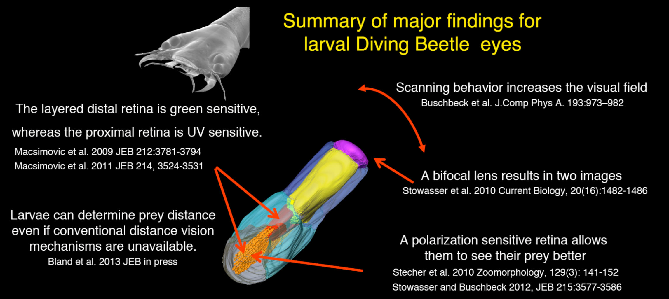
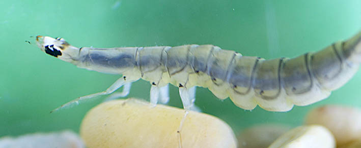
Although we continue to investigate Strepesiptera (see James et al 2016), in the past few years we have focused primarily on the functional organization of the principal eyes of the Sunburst Diving Beetle larvae (Thermonectus marmoratus), which are among the most bizarre and strangely organized eyes in the animal kingdom. Larvae use these eyes while they hunt their prey, and many of their eye specializations allow them to be highly effective visually guided predators. Research in our laboratory along these lines led to a series of papers including the discovery that they have multiple layered retinas, that one of those retinas expresses a green opsin whereas the other expresses a UV opsin, that the ultrastructure of the proximal retina supports polarization sensitivity, that larvae use scanning movements to track their prey, and perhaps among our most exciting findings, that in contrast to all other known eyes of the extant animal kingdom, their visual system operates with truly bifocal lenses. In addition we found that T. marmoratus larvae likely have evolved a novel way to assess object distances and some of our findings have already inspired new technologies. To study the function of these eyes our investigations range from light and ultrastructural microscopy, to optical measurements, intra – and extracellular electrophysiology and behavioral paradigms.
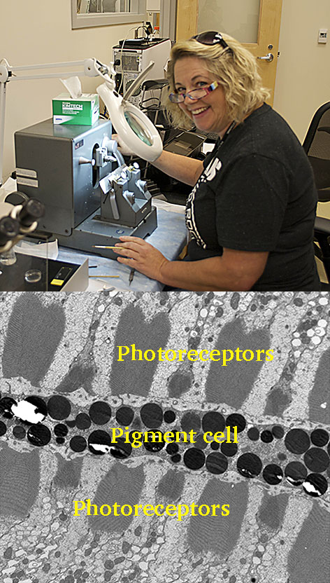
One of our current projects involves an Electron Microscopical investigation of photoreceptor recycling in diving beetle larval eyes. To maintain proper function, the membrane of photoreceptors needs to recycle. In vertebrates this is done with the help of the retinal pigment epithelium. However, in most invertebrates it remains unclear how this is done, and in many invertebrate eyes no pigment cells are near the photoreceptors. Our studies suggest that in diving beetle larvae there is a pigment cell adjacent to photoreceptors, and our preliminary results so far point towards the possibility that this cell is taking an active role in the photoreceptor-membrane recycling process. We are currently investigating photoreceptor recycling throughout the day and use toxins and fluorophores to investigate parallels in photoreceptor stack recycling between diving beetle larvae and vertebrates.
Lab members primarily associated with these projects: Annette Stowasser (former post-doc and ongoing collaborator), Rose Conley.

This project was funded by the National Science Foundation under Grants IOB-545978 (CAREER) and IOS-1050754 to Dr. Elke Buschbeck.
